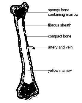
Long Bone With Diagram - Long Bones Medlineplus Medical Encyclopedia Image : Sectional diagram of a long bone.
byCarmella Parks-
0
Long Bone With Diagram - Long Bones Medlineplus Medical Encyclopedia Image : Sectional diagram of a long bone.. The carpals are connected to the five metacarpals that form the bones of the hand and connect to each of the fingers. A long bone consists of a long shaft (diaphysis) with two bulky ends or extremities (epiphyses) where articulation takes place. Helps keep bones light in weight. Its not option b because a fossa is a animal that is in the cat species. Knee synovial joint blank diagram.
Knee synovial joint blank diagram. Long bones contain yellow bone marrow and red bone marrow, which produce blood cells. These are strong bones because they must be able to withstand the force generated when though different long bones have different shapes and functions, they all have the same general structure. It is a long bone since its length is greater as compared to its width. Bone long diagram diaphysis tissue biology blood body cell compact humerus structure vector anatomical anatomy articular calcium cartilage detail education educational endosteum epiphysis forelimb health healthy human illustration joint long bone marrow medical medicine organ orthopedic.
Long Bone Anatomy Youtube from i.ytimg.com The carpals are connected to the five metacarpals that form the bones of the hand and connect to each of the fingers. A long bone is a drop from various monsters, usually those that drop big bones with some exceptions, at a universal rate of 1/400. The fishbone diagram dzone agile. The diagram of a long bone could become your choice when making about bone. A long bone is a bone that is significantly longer than it is wide. Each long bone has a shaft and two ends or extremities, which are usually articular. It is the only bone making up the upper arm. It is a long bone since its length is greater as compared to its width.
Bone long diagram diaphysis tissue biology blood body cell compact humerus structure vector anatomical anatomy articular calcium cartilage detail education educational endosteum epiphysis forelimb health healthy human illustration joint long bone marrow medical medicine organ orthopedic.
Also, they provide an environment for bone marrow, where the blood cells are created, and they act as a storage area for minerals, particularly calcium. Characterized by irregular spaces filled with red bone marrow that makes blood cells; Structure of the long bone with pictures learn with flashcards, games and more — for free. Simple foot diagram free wiring diagram for you, cattle bone diagram manual e books, bone classification and structure anatomy and physiology torso bone diagram data wiring diagram today. This is an online quiz called long bone diagram. Dimitrios mytilinaios md, phd there are five types of human bones: This is due to the shape of the bones, not their size. A long bone is a drop from various monsters, usually those that drop big bones with some exceptions, at a universal rate of 1/400. Human anatomy diagrams show internal organs, cells, systems, conditions, symptoms and sickness information and/or tips for healthy living. It is a long bone since its length is greater as compared to its width. They are one of five types of bones: This diagram depicts final long bone diagram. Download 3,638 diagram bone stock illustrations, vectors & clipart for free or amazingly low rates!
Start learning with our skeleton diagrams, bone labeling exercises and skeletal system quizzes! To know the architecture of compact and spongy (cancellous) bone. A long bone is a drop from various monsters, usually those that drop big bones with some exceptions, at a universal rate of 1/400. The tibia or commonly known as the shin bone is one of the bones you. Long bones contain yellow bone marrow and red bone marrow, which produce blood cells.
Human Being Anatomy Skeleton Structure Of A Long Bone Image Visual Dictionary from www.ikonet.com Long bones — a subtype of bones — are longer than they are wide. 12 photos of the diagram of long bone structure. Each system contains haversian canals surrounded by concentric lamellae of bone tissue 48. To recognise bone and understand its structure and to understand the processes by which bone can be formed. Its not option b because a fossa is a animal that is in the cat species. Human anatomy for muscle reproductive and skeleton. Long bone diagram blank human skeleton template. To know the structures of a synovial joint and a symphysis joint (intervertebral disc).
The articular surfaces are smooth, even after articular cartilage is removed.
Create your own flashcards or choose from millions created by other students. Found in the ends of long bones; Sectional diagram of a long bone. A long bone is a drop from various monsters, usually those that drop big bones with some exceptions, at a universal rate of 1/400. Human anatomy for muscle reproductive and skeleton. Dimitrios mytilinaios md, phd there are five types of human bones: The lower arm bones form the wrist joint with the carpals, a group of eight small bones that give added flexibility to the wrist. The outside of the flat bone consists of a layer of connective tissue called the periosteum. As shown in figure 2. Long bones, especially the femur and tibia, are subjected to most of the load during daily activities and they are crucial for skeletal mobility. Each system contains haversian canals surrounded by concentric lamellae of bone tissue 48. Knee synovial joint blank diagram. Being a homophone with the word the bone supports most of the major functions of the arm including lifting and throwing.
Structure of a long bone. The fishbone diagram dzone agile. When a human finishes growing these parts fuse together. Human anatomy for muscle reproductive and skeleton. Education chart of biology for immune system diagram in human being.
Anatomy And Physiology Of Animals The Skeleton Test Yourself Answers Wikibooks Open Books For An Open World from upload.wikimedia.org The fishbone diagram dzone agile. Long bones, especially the femur and tibia, are subjected to most of the load during daily activities and they are crucial for skeletal mobility. This is due to the shape of the bones, not their size. A long bone has • terminal portions of the bone with thinner cortices which consist largely of cancellous bone— these are the epihyseal regions forming the articulating parts of the bone, and the metaphyseal areas which link the epiphyseal and diaphyseal parts of the bone. 12 photos of the diagram of long bone structure. To know the architecture of compact and spongy (cancellous) bone. Anatomy of a long bone anna s anatomy websit. The shaft is also known as the diaphysis.
Sectional diagram of a long bone.
I'm not sure of what you mean by bone diagram. The lower arm bones form the wrist joint with the carpals, a group of eight small bones that give added flexibility to the wrist. Some bones in the fingers are classified as long bones, even though they are short in length. Tibia and fibula bone diagram. This diagram depicts final long bone diagram. The writing movement of the hands is also due to the presence and. These are strong bones because they must be able to withstand the force generated when though different long bones have different shapes and functions, they all have the same general structure. To know the architecture of compact and spongy (cancellous) bone. Each long bone has a shaft and two ends or extremities, which are usually articular. Each finger has three bones known as phalanges, except for the. The long bones are those that are longer than they are wide. It is a long bone since its length is greater as compared to its width. A long bone has • terminal portions of the bone with thinner cortices which consist largely of cancellous bone— these are the epihyseal regions forming the articulating parts of the bone, and the metaphyseal areas which link the epiphyseal and diaphyseal parts of the bone.